m-mode lv echocardiography plax | plax echocardiogram m-mode lv echocardiography plax VI. M-Mode Measurements This section provides guidance on selected M-mode measurements. VII. Color Doppler Imaging This section defines the basic imaging windows, display, and mea . Skursteņi, Augstākās kvalitātes keramiskie dūmvadi - malkas krāsnīm, kamīniem, visiem apkures katliem. 30 gadu garantija. Dūmvads PAR LABĀKO CENU!
0 · plax echocardiogram
1 · parasternal plax view echocardiogram
2 · m-mode of mv
The hotter the bath’s operating temperature, the faster and more severe this reaction will be. Very few fluids will hold up under these extreme conditions for more than a few days, however, Duratherm has created this line of heat transfer fluids specifically engineered for this challenging application
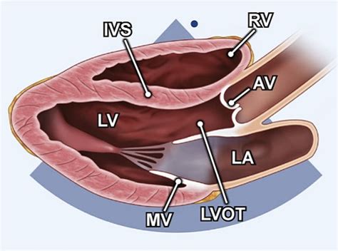
PLAX view. The parasternal long axis view (PLAX) is obtained with the transducer image marker directed toward the patient’s right ear and the sound beam directed to the spine. Slight . It is used to guide M-Mode echocardiography for left ventricular measurements. Initially the parasternal long axis view is obtained. When satisfactory images are available after .
How to measure the Ejection Fraction of left ventricle using m-mode or motion mode on parasternal long axis view.

THE AMERICAN SOCIETY OF ECHOCARDIOGRAPHY RECOMMENDATIONS FOR CARDIAC CHAMBER QUANTIFICATION IN ADULTS: A QUICK REFERENCE GUIDE FROM THE ASE .VI. M-Mode Measurements This section provides guidance on selected M-mode measurements. VII. Color Doppler Imaging This section defines the basic imaging windows, display, and mea .
Assessment of LV function with M-mode or 2-dimensional (2-D) echocardiography (Figure 2A) can be performed in the parasternal long- and short-axis views by .Use the zoom function in the PLAX view for optimal visualization of LV outflow tract (LVOT) and the aortic valve with visualization of AV cusp insertion points (annulus). Both MV leaflets and .
plax echocardiogram
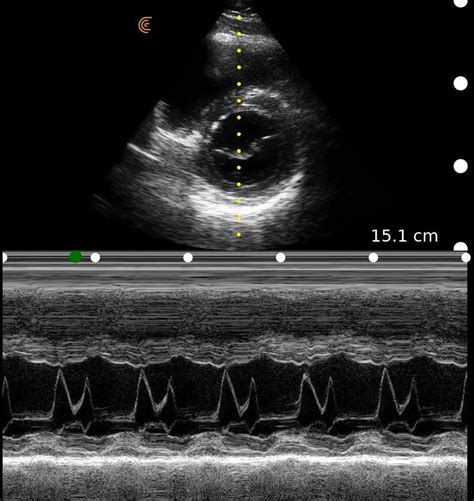
The M-mode from the LV at the mitral valve leaflet level may be useful to measurement: a) the diastolic inter-ventricular septum, b) the diastolic posterior wall of the LV. These may be .

PLAX M-mode: MV E-Septal separation (EPSS) EPSS is defined as the minimal distance between E point (most anterior motion of the AML during diastole) and a line tangential to the . M-mode imaging in the parasternal views will further elucidate mitral leaflet motion and define the duration of mitral-septal contact. Color Doppler and PW Doppler mapping should be integrated in the assessment of .
PLAX view. The parasternal long axis view (PLAX) is obtained with the transducer image marker directed toward the patient’s right ear and the sound beam directed to the spine. Slight adjustments in angle and rotation maybe necessary to demonstrate all .
It is used to guide M-Mode echocardiography for left ventricular measurements. Initially the parasternal long axis view is obtained. When satisfactory images are available after fine adjustments of the transducer position, M-Mode cursor is placed in such a .
parasternal plax view echocardiogram
How to measure the Ejection Fraction of left ventricle using m-mode or motion mode on parasternal long axis view.
THE AMERICAN SOCIETY OF ECHOCARDIOGRAPHY RECOMMENDATIONS FOR CARDIAC CHAMBER QUANTIFICATION IN ADULTS: A QUICK REFERENCE GUIDE FROM THE ASE WORKFLOW AND LAB MANAGEMENT TASK FORCE. Accurate and reproducible assessment of cardiac chamber size and function is essential for clinical care. A standardized methodology .VI. M-Mode Measurements This section provides guidance on selected M-mode measurements. VII. Color Doppler Imaging This section defines the basic imaging windows, display, and mea-surements for color Doppler imaging (CDI) to be integrated into the comprehensive transthoracic examination. Similarly, display of color
Assessment of LV function with M-mode or 2-dimensional (2-D) echocardiography (Figure 2A) can be performed in the parasternal long- and short-axis views by placing the calipers perpendicular to the ventricular long axis. Change in LV cavity dimensions during systole can be used to calculate LV fractional shortening and ejection fraction.Use the zoom function in the PLAX view for optimal visualization of LV outflow tract (LVOT) and the aortic valve with visualization of AV cusp insertion points (annulus). Both MV leaflets and 2 of the 3 aortic leaflets should be visible in good quality.
PLAX M-mode: MV E-Septal separation (EPSS) EPSS is defined as the minimal distance between E point (most anterior motion of the AML during diastole) and a line tangential to the most posterior excursion of the IVS within the same cardiac cycle M-mode imaging in the parasternal views will further elucidate mitral leaflet motion and define the duration of mitral-septal contact. Color Doppler and PW Doppler mapping should be integrated in the assessment of obstruction and, when present, determine the .
Rapidly moving structures such as the aortic valve and mitral valve, and endocardium have characteristic movements in M-mode. M-mode also has a great spatial resolution, which is useful for measuring ventricular dimensions in systole and diastole.PLAX view. The parasternal long axis view (PLAX) is obtained with the transducer image marker directed toward the patient’s right ear and the sound beam directed to the spine. Slight adjustments in angle and rotation maybe necessary to demonstrate all . It is used to guide M-Mode echocardiography for left ventricular measurements. Initially the parasternal long axis view is obtained. When satisfactory images are available after fine adjustments of the transducer position, M-Mode cursor is placed in such a .How to measure the Ejection Fraction of left ventricle using m-mode or motion mode on parasternal long axis view.
m-mode of mv
THE AMERICAN SOCIETY OF ECHOCARDIOGRAPHY RECOMMENDATIONS FOR CARDIAC CHAMBER QUANTIFICATION IN ADULTS: A QUICK REFERENCE GUIDE FROM THE ASE WORKFLOW AND LAB MANAGEMENT TASK FORCE. Accurate and reproducible assessment of cardiac chamber size and function is essential for clinical care. A standardized methodology .VI. M-Mode Measurements This section provides guidance on selected M-mode measurements. VII. Color Doppler Imaging This section defines the basic imaging windows, display, and mea-surements for color Doppler imaging (CDI) to be integrated into the comprehensive transthoracic examination. Similarly, display of color

Assessment of LV function with M-mode or 2-dimensional (2-D) echocardiography (Figure 2A) can be performed in the parasternal long- and short-axis views by placing the calipers perpendicular to the ventricular long axis. Change in LV cavity dimensions during systole can be used to calculate LV fractional shortening and ejection fraction.Use the zoom function in the PLAX view for optimal visualization of LV outflow tract (LVOT) and the aortic valve with visualization of AV cusp insertion points (annulus). Both MV leaflets and 2 of the 3 aortic leaflets should be visible in good quality.PLAX M-mode: MV E-Septal separation (EPSS) EPSS is defined as the minimal distance between E point (most anterior motion of the AML during diastole) and a line tangential to the most posterior excursion of the IVS within the same cardiac cycle M-mode imaging in the parasternal views will further elucidate mitral leaflet motion and define the duration of mitral-septal contact. Color Doppler and PW Doppler mapping should be integrated in the assessment of obstruction and, when present, determine the .
andrew mccarthy son versace
arredamento versace
All prices are the current market price. Dusknoir LV.X (Pokemon Japanese Intense Fight in the Destroyed Sky) prices are based on the historic sales. The prices shown are calculated using our proprietary algorithm. Historic sales data are completed sales with a buyer and a seller agreeing on a price.
m-mode lv echocardiography plax|plax echocardiogram


